

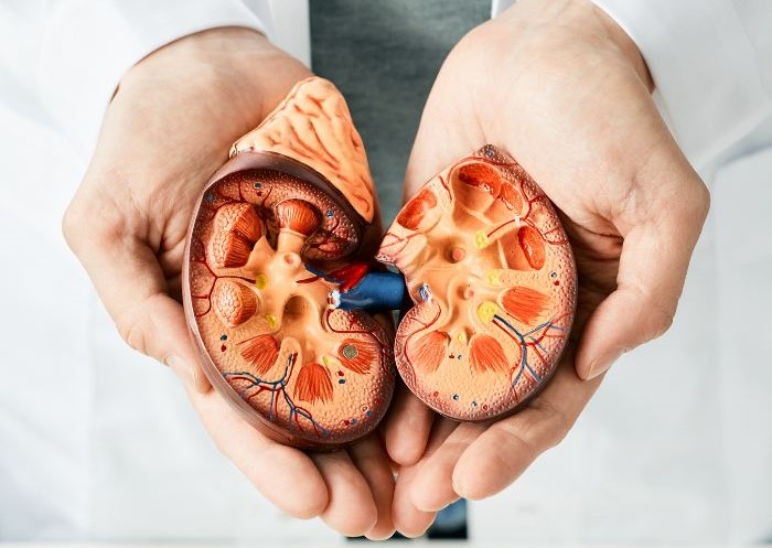
Finding a mass on a kidney scan can naturally cause concern, but it is important to remember that not every kidney lesion is cancerous. A kidney cyst is usually a fluid-filled sac, whereas a kidney tumour is an abnormal tissue growth that may be benign or malignant. Doctors rely mainly on imaging characteristics, rather than symptoms alone, to distinguish between the two. Anyone searching for the best urologist in Mumbai should expect a detailed review of the scan, medical history, kidney function, and personal risk factors before a treatment plan is considered.
Simple kidney cysts are common, particularly as people grow older, and most do not require treatment. Complex cysts need closer evaluation because they may contain thick walls, internal divisions, calcification, nodules, or solid areas. Kidney tumours are also frequently discovered by chance during an ultrasound, CT scan, or MRI performed for an unrelated concern. The best urologist in Mumbai will assess the complete picture instead of assuming that every cyst is harmless or every solid mass is cancerous. The next step depends on the lesion’s appearance, size, location, growth, symptoms, and the patient’s overall health.
A kidney cyst is a round or oval sac containing fluid. A simple cyst generally has a thin, smooth wall, clear fluid inside, and no solid components. According to the NIDDK, simple kidney cysts usually cause no symptoms and are often discovered during imaging performed for another reason.
Simple cysts are not the same as polycystic kidney disease. Polycystic kidney disease is an inherited condition in which numerous cysts develop and may gradually affect kidney function. Having one or two simple cysts does not mean someone has this genetic disorder.
Although most simple cysts remain harmless, a large cyst may occasionally cause pain, become infected, or obstruct the normal flow of urine.
A kidney tumour is a mass created by abnormal cell growth. It may be benign, meaning non-cancerous, or malignant. Renal cell carcinoma is the most common type of kidney cancer in adults, although an imaging scan alone may not always reveal the exact cell type.
Kidney tumours differ considerably in size, location, and behaviour. Some small masses grow slowly and can be monitored in selected patients. Others may need a biopsy, ablation, partial nephrectomy, or removal of the affected kidney.
Consulting the best urologist in Mumbai can help patients understand whether observation, additional imaging, biopsy, or treatment is appropriate. Decisions should not be made from one ultrasound report without considering the complete clinical picture.
Doctors look at several characteristics when distinguishing between a cyst and a tumour:
• Contents: Simple cysts contain fluid, while tumours usually contain solid tissue.
• Shape: Simple cysts are generally round with thin, smooth walls. Tumours may have uneven or irregular borders.
• Contrast enhancement: Solid tumours often show enhancement after contrast is administered. Simple cysts do not.
• Internal features: Thick walls, divisions, nodules, or combined solid and fluid areas make a lesion more complex.
• Cancer risk: Simple cysts are usually benign. Complex cysts and solid masses require further risk assessment.
• Management: Simple cysts may need no further care, whereas tumours may require monitoring, biopsy, ablation, or surgery.
A cystic lesion can sometimes represent cystic kidney cancer. Therefore, the words “cystic” and “benign” should not automatically be treated as having the same meaning.
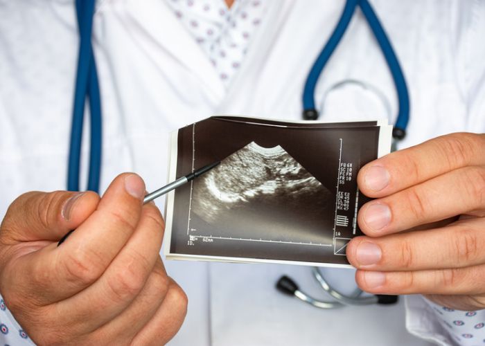
An ultrasound can often identify a straightforward fluid-filled cyst. However, a contrast-enhanced CT scan or MRI may be needed when the lesion has uncertain or complex features. Contrast imaging shows whether the walls, internal divisions, nodules, or solid areas demonstrate enhancement.
Doctors generally assess:
• The size and exact location of the lesion
• Whether it is fluid-filled, solid, or mixed
• Wall thickness and internal divisions
• Response to contrast
• Changes compared with previous scans
• Current kidney function
• Existing symptoms
• Family history and other risk factors
The Bosniak classification system is commonly used to categorise cystic kidney masses according to their imaging features. Lower categories are generally considered benign, while higher categories carry more concern and may require surveillance or treatment.
An unclear or indeterminate scan should be reviewed by the best urologist in Mumbai in consultation with an experienced radiologist. This combined assessment helps avoid both unnecessary treatment and delays in managing a potentially concerning lesion.
Many kidney cysts and tumours do not produce symptoms, particularly when they are small. Possible symptoms may include persistent pain in the side or back, fever, an abdominal lump, unexplained weight loss, or reduced appetite.
These symptoms can also occur with several non-cancerous conditions and do not confirm a kidney tumour. Severe pain, fever with chills, vomiting, or difficulty passing urine requires prompt medical assessment.
A simple cyst that is not causing symptoms usually requires no treatment. If a cyst becomes large, painful, infected, or obstructive, it may be drained and treated to reduce the chance of recurrence. Surgical removal may be considered in selected cases.
Complex cysts may require scheduled follow-up scans or surgery, depending on their Bosniak category, size, imaging appearance, and changes over time.
Treatment options for kidney tumours may include active surveillance for selected small masses, thermal ablation, partial nephrectomy, or radical nephrectomy.
A partial nephrectomy removes the tumour while preserving the healthy portion of the kidney. Radical nephrectomy removes the entire affected kidney and may be required for certain larger or more complex tumours. The National Cancer Institute explains that treatment decisions depend on factors such as cancer stage, age, general health, and individual circumstances.
The best urologist in Mumbai should explain the advantages and limitations of every available option, including its possible effect on kidney function, recovery, and future monitoring.
1. Can a kidney cyst turn into cancer?
A simple kidney cyst does not usually become cancerous. However, complex cystic masses may have malignant potential and need proper imaging assessment.
2. How can doctors tell a cyst from a tumour?
Ultrasound, contrast-enhanced CT, and MRI can evaluate fluid content, solid tissue, wall thickness, internal nodules, and contrast enhancement.
3. Does every kidney tumour require surgery?
No. Selected small masses may be monitored, biopsied, or treated with ablation. Management depends on their size, appearance, growth, location, and the patient’s health.
4. When should I consult the best urologist in Mumbai?
Consult a urologist when imaging shows a complex cyst, solid mass, growing lesion, uncertain diagnosis, persistent symptoms, or changes in kidney function.
5. Is a kidney biopsy always required?
No. A biopsy can be helpful in selected cases, but imaging and clinical findings may sometimes provide enough information to plan treatment.
6. Can a kidney tumour be removed without removing the kidney?
Yes. Partial nephrectomy removes the tumour while preserving healthy kidney tissue. Whether it is suitable depends on the tumour’s size, position, and complexity.
7. How often should a complex kidney cyst be monitored?
The follow-up schedule depends on the Bosniak category, lesion stability, age, kidney function, and general health. The treating specialist will recommend an appropriate timeline.


Dr. Mangesh Patil is a renowned Urologist, and is trained in Endourology and Robotic Surgery , with professional experience of more than 10 years .
© 2024 – Urocare Clinic. All Right Reserved | Maintained By Rebecca Digital